Clinic for the congenial malformation of hand & foot
Polydactyly

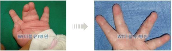
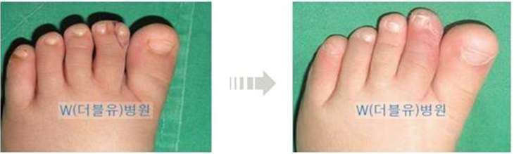
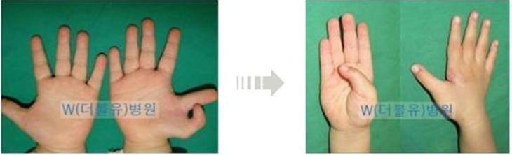
Polydactyly refers to the condition of more than6 fingers or toes. The most frequent symptom is the formation of another finger on the thumb or the little finger. In the case of the Polydactyly of the thumb, the extent and the range of the surgery differ very much due to extensive range of configuration, arrangement and sizes of the bones, ligament and tissues.

In the case of the Polydactyly that occurs on the thumb, past family history accounts for only 10% of the cases and it is deemed to occur sporadically. Therefore, it is deemed that there need not be serious concerns for the genetic cause of Polydactyly.

In simple cases in which only the tissues are attached, surgical procedure can be performed easily. However, if bones, joints and ligaments are connected complexly, it becomes a difficult and complicated surgical procedure that requires several surgical procedures to achieve normal appearances and functions of the fingers. Therefore, efforts need to be put in to prevent such malformation at the time of the first surgery.

1. Time of the surgery
In the modern medicine, it is the widely accepted theory that it is advisable to perform surgery on congenital malformation as early as possible. Moreover, parents wish to illustrate normal hands of the infant to the relatives and friends at the 100-day or 1st birthday party. Accordingly, they request the surgery be performed prior to the 100th day of the birth of the infant. In the case of simple Polydactyly, it is possible to perform the surgery prior to the 100th day of the birth. However, if the problems of the bones, joints and ligaments are accompanied, it is advisable to perform the surgery at around 1 year from the birth. Accordingly, there is a trend of undergoing primary surgery at about 8~10 months after birth nowadays.
2. Anesthesia and surgical technique
In simple cases, it is possible to perform excision simple sedation anesthesia. The condition of the infant prior to the surgery becomes more important earlier the surgery is performed. It is advisable to postpone the surgery by about 2 weeks if there are symptoms of common cold in the infant. Sedation anesthesia, unlike the general anesthesia, refers to the performing the surgery by injecting localized anesthesia in the hand while the infant is in sedated conscious state. When the infant is fully awake after the surgery, the infant can be taken home immediately. However, in the case of the surgery that takes long time with the need to work on the joints, ligaments and bones, general anesthesia is necessary along with 1~2 days of hospitalization.

The surgical techniques can be categorized largely into the following simple excision procedure, reconstruction procedure following excision and Bilhaut-Cloquet procedure.
A. Simple excision procedure
If only the tissues are attached, it is possible to remove the unnecessary tissues by using string or devices immediately after the birth. However, if bone structure is involved, it is possible to perform excision surgery at about 5~6 months after birth at which sedation anesthesia can be implemented.
B. Reconstruction procedure following excision
In general, if the fingernail size of the polydactyly finger becomes 70~80% of that of the normal finger, the remaining finger is excised. At this time, excision is made by ensuring that the necessary tissues are preserved rather than simply cutting off the excess finger because it is necessary to improve the appearance and function of the finger by suturing the tissues of the surplus finger to be excised with the tissues of the finger left intact if the bones, joints and ligaments are interconnected. In majority of the cases, the joint needs to be reconstructed since the ligaments that form the joints of the finger are weak. Rarely, osteotomy of portion of the bone is necessary as well.
C. Bilhaut-Cloquet procedure
If 2 polydactyly fingers have similar size and neither has fingernail size that is 70% of that on normal finger at the maximum, surgical technique of combining the 2 fingers to create a single finger is performed. After having appropriately cut the bones and joints of the both fingers, bones are fixated and the ligaments are connected. However, in this case, 2 fingernails are combined to make one fingernail, there is the problem of the split in the fingernail as it grows and the reconstructed joint would have deteriorated functions.
3. Complications: Secondary corrective procedure
Simple complication that is observed commonly after the primary surgery include bulging of portion of the bone with the regrowth of the bone due to the failure to completely remove the growth point of the bone in the excised finger. In such case, it is possible to make correction by excising the bone and the surrounding tissues that are bulging out.

However, in the case of polydactyly thumb, it grows in Y-shape from birth and will not grow in straight line even if one of the axes is cut off. Just as one of the axes of Y is cut off, the remaining portion does not become straight, the formation of deformation due to the bending of the joint after the primary surgery is referred to as the 'Z' shaped deformation of the finger (angle formation).

Although the extent of the results following the surgery in the case of Polydactyly with complicated configuration differs substantially, it is the most common complication that does not fail to manifest. Accordingly, it would not be excessive to emphasize the importance of the primary surgery repeatedly for this reason. Therefore, maximum efforts need to be put in to prevent this deformation at the time of the primary surgery. Although it differs amongst various scholars, a surgical procedure can be performed at about the age of 5~6 years old if the deformation is severe or corrective surgery is performed by implementing osteotomy at the time of completion of the growth of the bones, that is, at about the time of graduation from elementary school. It is not easy to obtain fully satisfactory results from a single execution of such surgical procedure.
4. Care after the surgery
In most cases, the suture threads are removed after about 2 weeks, and the area of the surgery can be washed with water thereafter and it is advisable to frequently massage the affected area by using ointment that minimizes formation of scars. In the event of having performed replantating of the ligament for joint reconstruction, it would be advisable to apply cast for about 4 weeks. In addition, it is possible to assist proper growth of the thumb by applying small splint to the hand occasionally in order to prevent secondary Z-deformation.

In conclusion, since the thumb is an important finger that accounts for more than 50% of the functions of the hand, efforts must be put into maintain the anatomical configuration and functions of the thumb as close to the normal condition as possible if there is congenital malformation. For this purpose, the parents need to ponder over when and how to apply surgery from the position of the infant rather than themselves, and the surgeon needs to put utmost efforts in fully reconstructing the functions of the hand without considering it as a simple surgery of excising the surplus tissues.
Syndactyly

It refers to the condition of failure of the finger to be properly separated with 2 or more fingers fused together. The skin between the fingers has the appearance of webbed feet of ducts. It is the malformation of the fingers with the highest prevalence rate among all the congenital malformation of fingers along with the Polydactyly. It is relatively common to find such malformation on both hands simultaneously and symmetric occurrence is not that rare either. Such malformation is accompanied in the toes as well.

1. Embryology
Observation of the formation of finger during the embryonic stage illustrates formation of 5 ridges at the hand plates at about 6 weeks that will become the fingers. This is referred to as the digital bud. Although these digital buds need to grow out of the hand plate and become separated normally at about 6~8 weeks of formation of embryo, if they fail to be separated, syndactyly will result.
2. Cause and frequency
Only various causes have been presumed without fully pinpointing the accurate cause. There is an assertion that the differentiation failure in separation of fingers (toes) due to the embryonic extinction of the cells planned at about 6~8 weeks is the cause of this phenomenon. It is most common that the third and the fourth fingers are attached in the hand while the second and the third toes are attached in the foot. There are cases in which only the skin is attached while there also are cases in which even the bones are fused together. In approximately 1/3 of the cases, accompanied malformation manifests with highly diverse types. It is one of the most common congenital malformations of the hands with the average prevalence rate of 1 in 6,000-8,000 people and more common in male than female. In general, approximately 15% of the patients have past family history.
3. Classification
It is categorized into complete and incomplete types depending on the extent of the fusion of the soft tissues. If even the fingernails are fused together, then it is a complete type while the incomplete type has up to the middle of the fingers fused. It is also categorized into simple and complex type depending on the types of the tissues that are fused together. Accordingly, although it would be necessary to take radiologic image of the fingers to be certain, cases in which the skins are fused is classified as the simple type while complex interconnection of the bones is referred to as complex type. In such case, the phenomenon is accompanied by abnormality in the nerves, blood vessels and tendons extended into each of the fingers.
4. Time of treatment and surgical technique
The time of the surgery differs depending on the complexity and relevant areas of the malformation. Although it is advisable to perform the surgery before the infant reaches the age of 1 in most cases, there are cases in which surgical procedure is postponed until the age of 2 by which the functional characteristics of the hand can be ascertained. Similar to Polydactyly, there also are many cases in which one would have to wait for the recovery of the functions. If the time of the surgery is delayed, there may be deformation of bones. For example, if the fourth and the fifth fingers are fused, the 2 fingers with difference in the lengths will induce the deformation of the bent bones of both fingers if they stay fused for prolonged period of time.

The time of the surgery differs depending on the complexity and relevant areas of the malformation. Although it is advisable to perform the surgery before the infant reaches the age of 1 in most cases, there are cases in which surgical procedure is postponed until the age of 2 by which the functional characteristics of the hand can be ascertained. Similar to Polydactyly, there also are many cases in which one would have to wait for the recovery of the functions. If the time of the surgery is delayed, there may be deformation of bones. For example, if the fourth and the fifth fingers are fused, the 2 fingers with difference in the lengths will induce the deformation of the bent bones of both fingers if they stay fused for prolonged period of time.

Hospitalization is necessary since the surgery is conducted under general anesthesia and may take approximately 2~3 days to as much as 5~6 days in the case of severe cases. The time taken for the actual surgery tends to be relative long due to the extensive need for suturing because of the skin graft involved, unlike Polydactyly. The suture thread can be removed in about 2 weeks. Obviously, it is covered by the medical insurance like any other congenital malformations. Although it is possible to separate the fingers that are fused at more than 2 locations in a single surgical procedure, it is advisable to perform 2~3 surgical procedures in most cases.

The purpose of the surgery is to separate the fused area to results in fully independent fingers with appropriate space between the fingers and preservation of the original functions of the fingers. It is also important to prevent the contracture that occurs as the fingers grow. In the case of even the fingernails being fused, creation of the tissues and skins around the fingernails of the both fingertips is very complicated and difficult.

The surgical procedure involves separating of the fingers in zig-zag format rather than simply making division between the fingers in order to prevent the occurrence of the secondary contracture of the skins later on.

The areas with insufficient skin must be supplemented through skin graft after the separation of the fingers.

Although skins from the groin or buttock area were grafted frequently in the past, the color and the texture of the skin differs substantially from those of the fingers, and the scars resulting from the areas from which the skin was take were substantial. Accordingly, skin from the sole of the foot skin is used for the skin graft in order to overcome such disadvantages of the skin from groin or buttock nowadays. Some of the advantages of the skin from the sole of the foot include texture similar to that of the finger and lesser extent of the secondary contracture. Furthermore, since the sole of the foot has thick skin, it has the advantages of leaving little scar even after skin has been taken off and not exposing the scar easily.
5. Post-surgical care and secondary surgery
The suture thread is taken out after 2 weeks of the surgery in most cases, and washing the affected area with soap water and massaging with ointment are necessary thereafter. Continuous massage is necessary in order to reduce scars and to make the skin softer since the skin has been grafted. Moreover, it is recommendable to use the ointment that minimizes the scar. Massage using ointment is also necessary for the area of the sole of the foot from which the skin was taken off once the wound is healed.

Since the incision line left behind on the finger and the scar after the skin graft do not just remain inactive but rather are subjected to continuous pulling force, contracture of the skin will occur. Therefore, the designing of the incisions at the time of the surgery is made in zig zag shape rather than in a straight line. Even with such incision line, the space between the separated fingers either fuse again or narrows and contracture manifests along the incision line, thereby frequently necessitating secondary surgery.

Secondary surgery can be executed after a minimum of 6~12 months after the initial surgery. It can be performed relatively simply unlike the initial surgery. Such secondary surgery may be needed 1~2 times after the primary surgery.

In addition, deformation of the bending of the bones sideways may be accompanies in syndactyly. In such case, osteotomy needs to be performed at the bent section of the bone during the secondary surgery in order to straighten the bones. If the bones are fixated with wire, there is no need for cast and there may be approximately 4~6 weeks of treatment necessary depending of the progress of the fusion of the bones following the corrective surgery.
Polysyndactyly

Polysyndactyly refers to the concurrent occurrence of Polydactyly in which more than 6 fingers or toes exist and syndactyly in which the fingers or toes are not fully separated but 2 or more have been fused together (with the appearance of webbed skin between the fingers or toes like those of the duck).